X-ray absorption spectroscopy (XAS) is a technique for probing the local electronic structure of materials. Due to the need for a high signal-to-noise ratio, high brilliance source, and tunable frequency range, synchrotron X-ray radiation is generally used to analyse the small amount of material comprising a molecular monolayer.
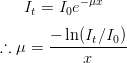
During an XAS measurement, a monochromated X-ray beam is tuned to an energy range where core electrons of the sample can be excited and passed through a sample. This excitation is observed as a loss in intensity of the transmitted X-ray beam, and the difference in intensity between the incident beam (I0) and transmitted beam (It) is expressed as the absorption coefficient μ:
Due to the complex nature of the interaction between the incident X-rays and the crystal, the resulting XAS spectra exhibit several distinct features, as shown in Figure 2.11.
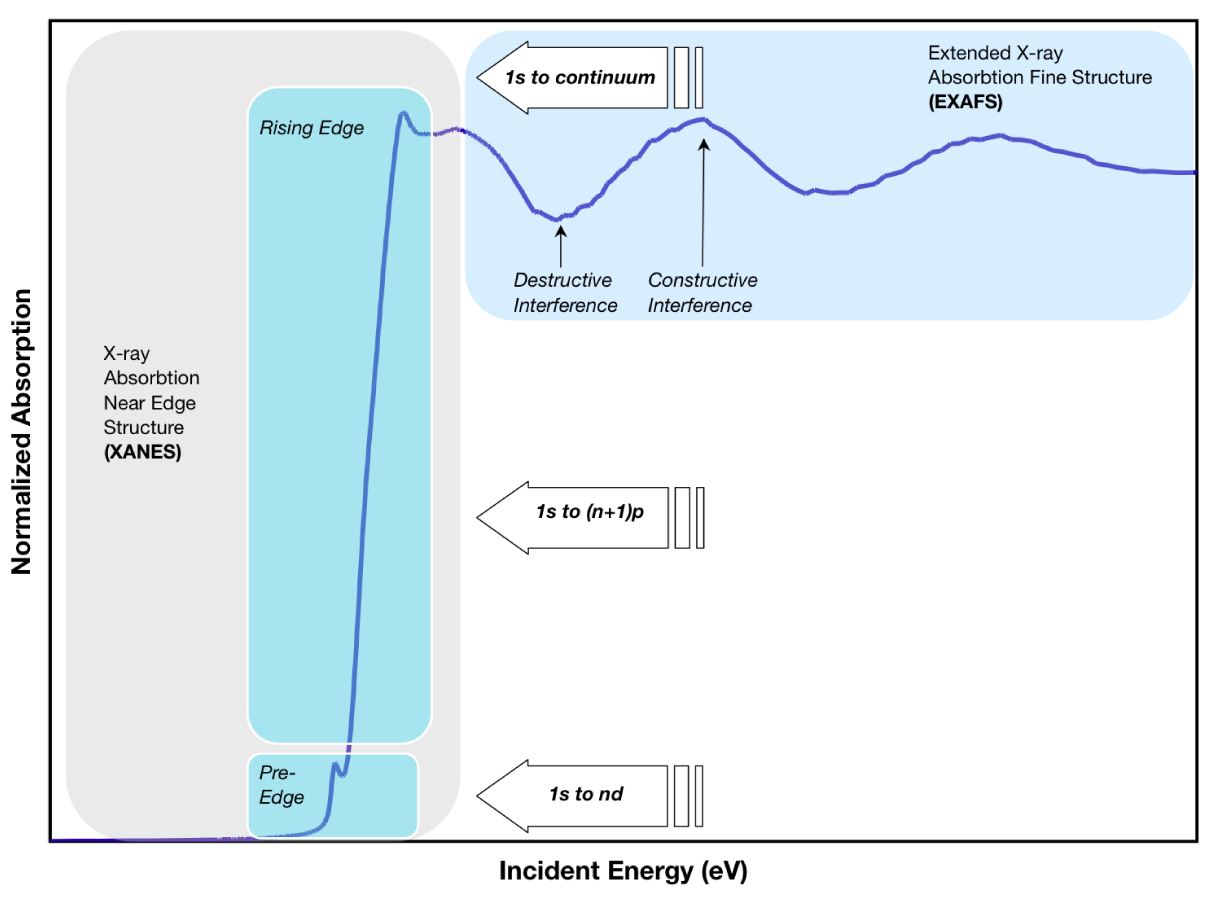
Walther Kossel first explained the structure of X-ray absorption peaks as due to electronic transitions to the first unoccupied molecular orbital above the Fermi energy (chemical potential). In turn, these were named the “Kossel structure”, however the more usual name for this region today is the absorption edge region.
The dominant feature of the spectrum produced by X-ray absorption is the “rising edge”, which occurs at the core-level binding energy of the absorbing atoms, with some smaller “pre-edge” features present at lower energies. Measurements of this are usually referred to as XANES (X-ray Absorption Near-Edge Structure) or NEXAFS (Near-edge X-ray Absorption Fine Structure). Mn 2p NEXAFS spectra are presented in Chapter 5 (Figure 5.7).
The oscillatory structure which can extend for hundreds of eV past the absorption edge was explained by Ralph Kronig, who had earlier first postulated the idea of electron spin. This so-called “Kronig structure” was attributed to multiple scatterings of the excited electron by the crystal lattice, and Kronig’s equation formed the basis for many early publications on X-ray absorption, although his theoretical basis was somewhat flawed. It was not until the 1970s when Edward Stern and colleagues at the University of Washington and Boeing Scientific Research Laboratory teased out the details of the theory [58]. The modern description for the high-energy structure is EXAFS (Extended X-ray Absorption Fine Structure).
At higher energies, scattering of the excited photo-electron from neighbouring atoms gives rise to a fine structure (EXAFS) due to the constructive and destructive interference of the excited and scattered electrons. When NEXAFS (XANES) and EXAFS are combined, the overall technique is known as XAFS (X-ray absorption fine structure).
The edges are designated according to the principal quantum number of the core electron excited; n = 1, 2, and 3 correspond to the K-, L- and M-edges, respectively. These transitions are shown schematically in Figure 2.12
![]()
Since a photo-electron is excited into an unoccupied orbital or band, XANES (NEXAFS) primarily gives information on the sample’s unoccupied states in the presence of the core-hole induced by the initial excitation from the core-level, and thus the final NEXAFS state can be thought of as a core “exciton”.